PRKAG2 Cardiomyopathy: A Case-Control Study on the Diagnostic Yield Of Histopathology and Ultrastructural Analysis from Endomyocardial Biopsy
Abstract
Background
The histopathological features of PRKAG2 cardiomyopathy have been reported in a fragmentary manner.
Objective
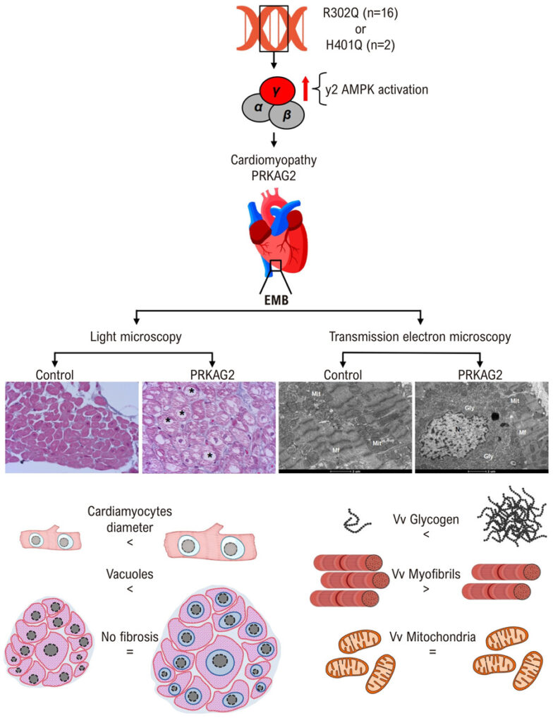
We aimed to systematically evaluate the cardiac pathological features of PRKAG2 cardiomyopathy in a large patient cohort and assess their diagnostic potential compared to genetic sequencing.
Methods
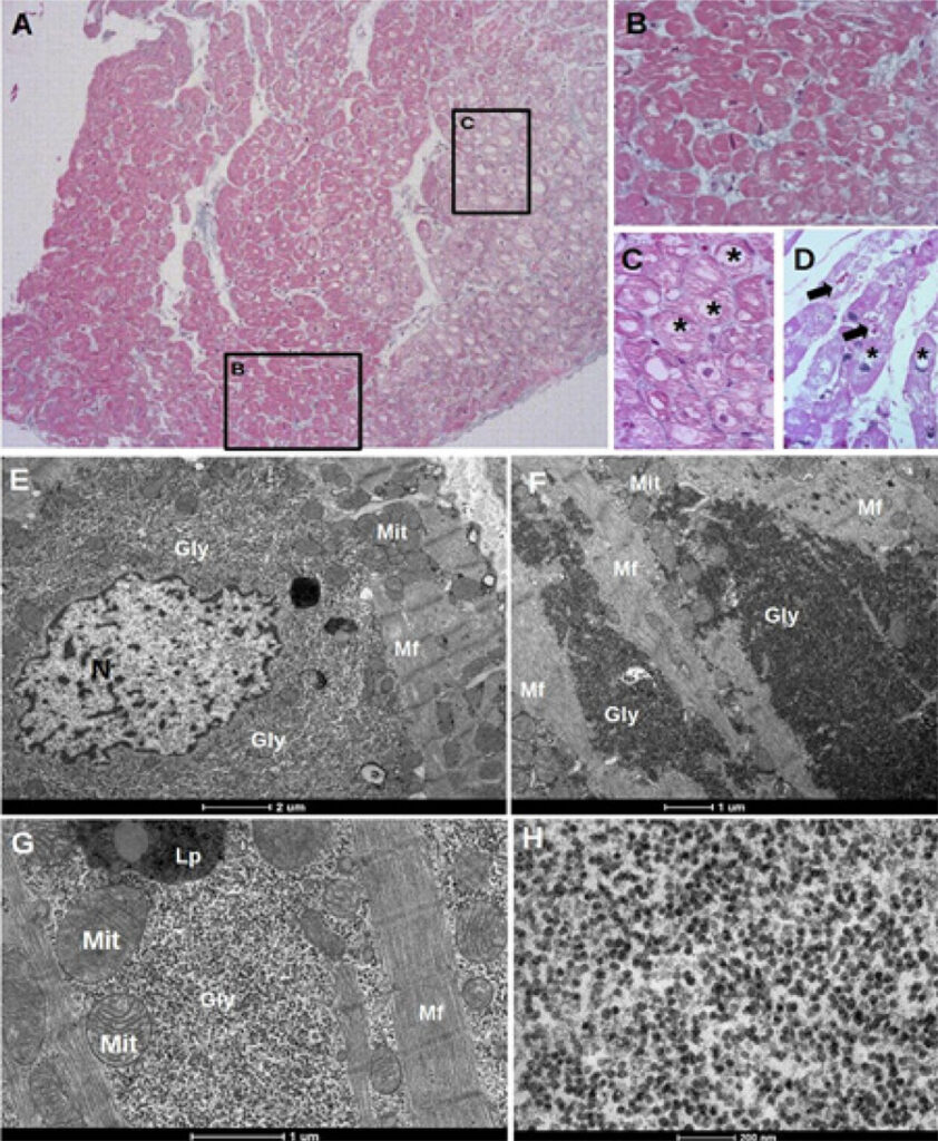
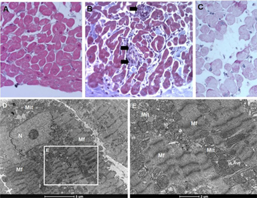
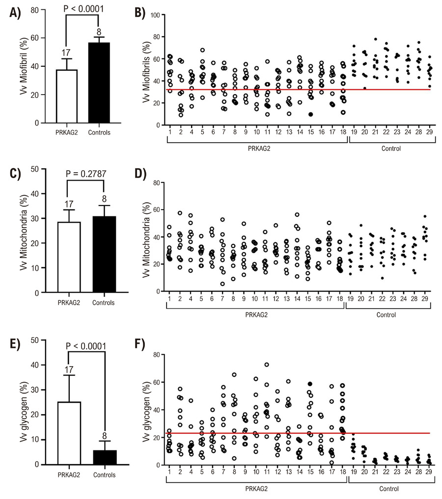
We conducted an observational, cross-sectional, case-control study including 18 patients with PRKAG2 cardiomyopathy and 11 heart transplant recipients as controls. All patients underwent percutaneous right ventricular endomyocardial biopsy. Tissue samples were analyzed using H&E staining, Periodic Acid-Schiff staining for glycogen, Masson’s trichrome for fibrosis, and ultrastructural assessment by transmission electron microscopy. Statistical significance was set at p < 0.05 for all analyses.
Results
PRKAG2 cardiomyopathy hearts exhibited significant cardiomyocyte enlargement, normal-appearing mitochondria, extensive vacuolization of most myofibers, minimal interstitial fibrosis (only two patients had mild fibrosis), and no inflammatory cell infiltration. Transmission electron microscopy revealed abundant cytosolic glycogen, primarily in the perinuclear region, with additional deposits in intermyofibrillar and subsarcolemmal areas. This pronounced glycogen accumulation, consistently observed in all PRKAG2 patients, was absent in controls.
Conclusion
Histological and ultrastructural examination of right ventricular endomyocardial biopsy samples reveals a distinct set of features that strongly suggest PRKAG2 cardiomyopathy.
Images
Image gallery fullscreen viewer
Comments are moderated by the editorial team before publication. Discussions should be evidence-based and use scientific language. Conflicts of interest should be declared.