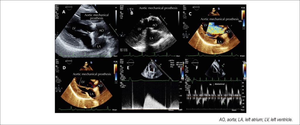
Transthoracic echocardiography (A) and transesophageal echocardiography (B) images showing fixed (arrows) hyper-refringent sub- and supra-prosthetic images suggestive of pannus. Color flow mapping demonstrates significant aortic prosthetic regurgitation (C) with no signs of prosthetic regurgitation in the next cardiac cycle (D) on the same projection. Aortic prosthetic regurgitating jet with a pressure half-time of less than 200 msec (E). Presence of intermittent holodiastolic reversal flow in the abdominal aorta (F; arrows).
Comments are moderated by the editorial team before publication. Discussions should be evidence-based and use scientific language. Conflicts of interest should be declared.
Comments are moderated by the editorial team before publication. Discussions should be evidence-based and use scientific language. Conflicts of interest should be declared.