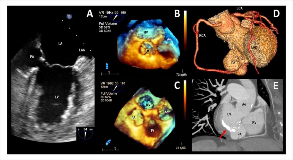
A. Transesophageal echocardiography (TEE) showing a pulsatile perivalvular echo-free space of 32 × 23 mm corresponding with a basal inferior pseudoaneurysm (PA). B, C. Real-time 3D TEE showing PA and its relationship with adjacent structures. D. A 3D volume-rendered cardiac CT angiogram showing PA and coronary arteries. E. Maximum intensity pixel projection reconstruction using a short axis view at the level of the mitral valve showing PA. Note the severe mitral annular calcification (arrow).
Comments are moderated by the editorial team before publication. Discussions should be evidence-based and use scientific language. Conflicts of interest should be declared.
Comments are moderated by the editorial team before publication. Discussions should be evidence-based and use scientific language. Conflicts of interest should be declared.