Simultaneous Dual Coronary Fistulas
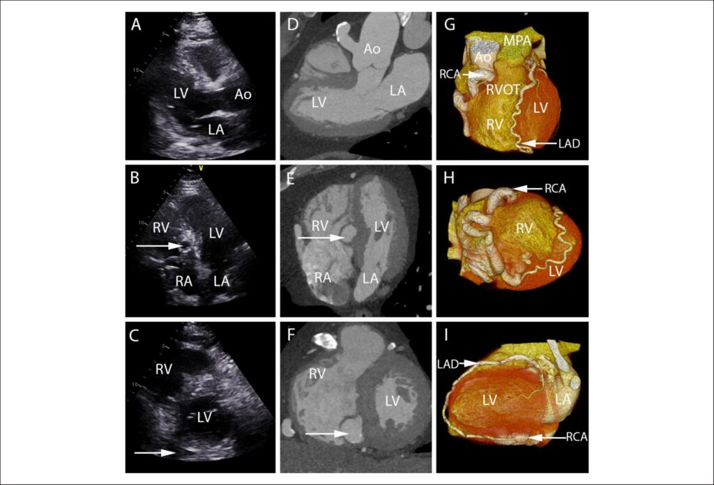
A 61-year-old man with type II diabetes mellitus was referred with breathlessness on exertion. On auscultation, there was a continuous ejection systolic murmur on the left upper sternal border. Transthoracic echocardiography showed a dilated vessel in aortic wall in the parasternal long axis view () and a spherical lesion in the apical 4-chamber view (). A coronary computed tomographic angiographic (CTCA) study revealed a dilated and ectatic right coronary artery (RCA). It arose from the ascending aorta at the 12 o’clock position and followed a tortuous course around the right sided atrioventricular groove before passing into the basal inferoseptum draining into the base of the right ventricle. An additional fistulous connection could be detected between the posterior descending artery of the RCA and the left anterior descending artery (LAD-RCA fistula) (, , ). After a normal dobutamine stress echocardiogram, a decision for continued medical therapy was taken.
[…]
Images
Image gallery fullscreen viewer
Comments are moderated by the editorial team before publication. Discussions should be evidence-based and use scientific language. Conflicts of interest should be declared.