Large Lymphoma Involving the Aortic Arch and its Branches: Demonstration by Echocardiography and Contribution to the Assessment of Hemodynamic Repercussions
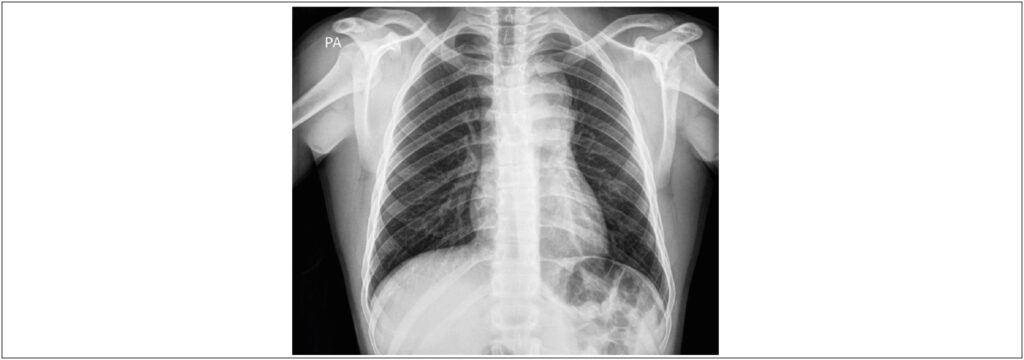
Chest X-ray in posteroanterior incidence demonstrating enlargement of the upper part of the mediastinus.
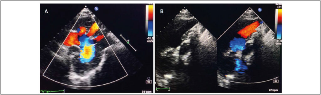
A – Transthoracic echocardiography (suprasternal notch view) showing color Doppler flow of the aortic arch (and its main branches) and proximal descending aorta; B – Two-dimensional image and color Doppler flow of the brachycephalic trunk.
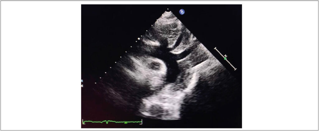
Transthoracic echocardiography (suprasternal notch view) showing a mass of heterogeneous echogenicity involving the aortic arch (and its main branches) and the proximal descending aorta.
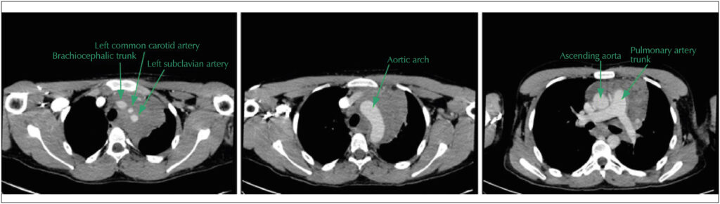
Contrast-enhanced chest computed tomography revealing extensive solid mediastinal tumor involving pulmonary artery, aortic arch (and main branches) and proximal descending aorta. There is no evidence of compression or invasion.
Comments are moderated by the editorial team before publication. Discussions should be evidence-based and use scientific language. Conflicts of interest should be declared.