Spontaneous Pneumomediastinum
Case
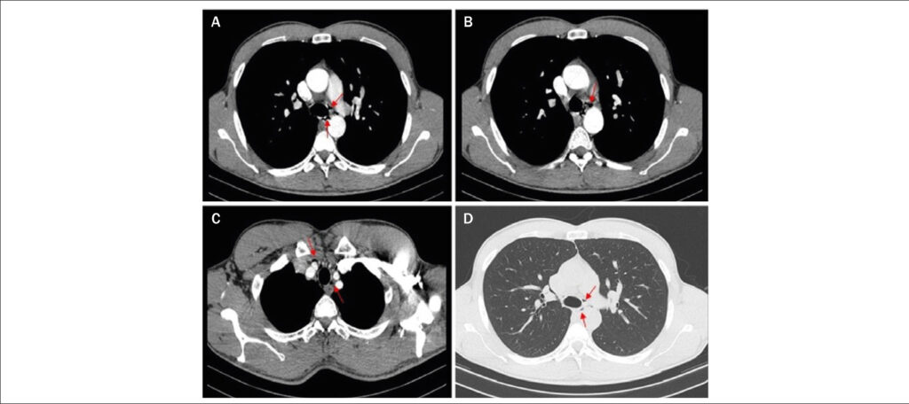
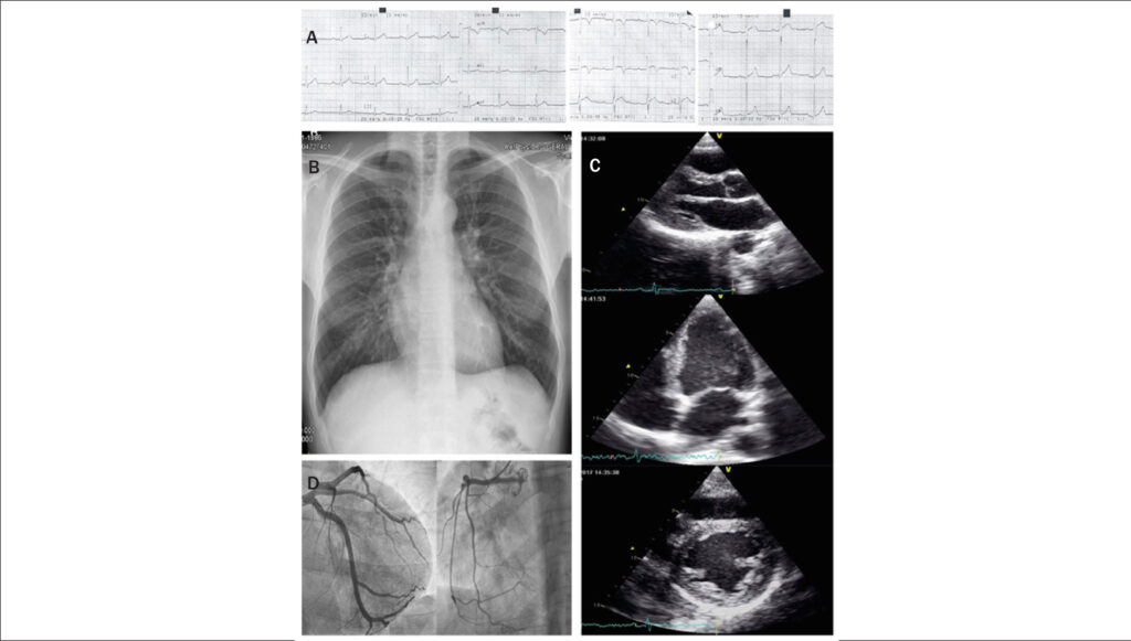
A 31-year-old male athlete presented with acute chest pain that was aggravated with swallowing after completing a marathon. Physical examination findings and his vital signs were normal. Electrocardiography revealed showed sinus rhythm, an incomplete right bundle block, and a negative T wave in V1-V2. A blood analysis showed normal D-dimer values and a slightly elevated troponin-I level (maximum at 72 hours: 0.52 ng/mL; normal, <0.07 ng/mL) with fluctuations. Chest X-ray, echocardiography, and coronary angiography findings were normal. Cardiac computed tomography (CT) revealed small gas collections on the superior and middle mediastinum compatible with spontaneous pneumomediastinum (SP). ( and ) SP is rare, usually benign, and often underdiagnosed. Intense physical exercise is a recognized cause. Cardiac CT assesses the extension, causative factors, and pathologies and discloses the diagnosis of SP when chest X-ray findings are normal.
[…]
Images
Image gallery fullscreen viewer
Comments are moderated by the editorial team before publication. Discussions should be evidence-based and use scientific language. Conflicts of interest should be declared.