Partial Prosthetic Mitral Valve Dehiscence: Transapical Percutaneous Closure
An 80-year-old woman with a history of mitral and aortic prosthesis replacement with biological prostheses due to endocarditis presented worsening dyspnea. A transthoracic echocardiogram demonstrated a paravalvular regurgitation between the left ventricle and left atrial appendage. Given her high-risk surgery (EuroSCORE-II: 38%), a percutaneous approach was performed for definitive closure.
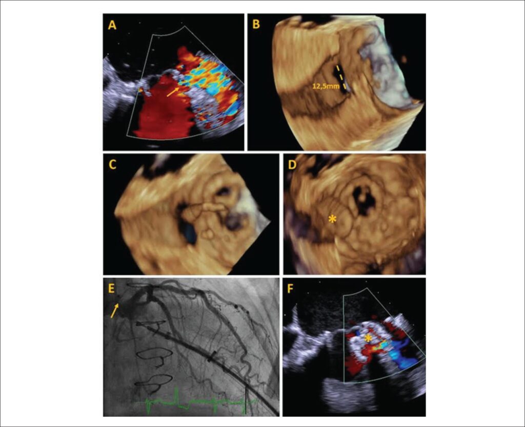
Transesophageal echocardiography (TEE) peri-procedure allowed the visualization of a partial dehiscence of the mitral prosthesis (Panel A, ). Through the 3D images, a tunneled defect with wall dissection measuring 12.5 mm of maximum diameter (Panel B, ) was observed. Using a transapical pathway and collecting three-dimensional (3D) images in real time, a 12 mm Amplatzer septal prosthesis was positioned, occluding the entire defect. The TEE 3D image demonstrated savings of adjacent structures and absence of pericardial effusion during closure. Coronary angiography demonstrated no arterial compromise. A slight residual flow was detected after device implantation (C-F Panels ).
[…]
Images
Image gallery fullscreen viewer
Comments are moderated by the editorial team before publication. Discussions should be evidence-based and use scientific language. Conflicts of interest should be declared.