Comparison between right and left ventricular myocardia during the human fetal period. Stereological evaluation
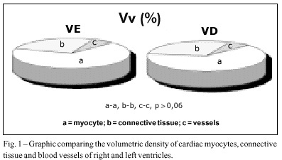
PURPOSE: To develop a stereological comparison between right (RV) and left ventricle (LV) myocardium during the third human gestational trimester. METHODS: Five human fetal hearts of the third trimester provided representative samples of 5 RV myocardium and 4 LV myocardium. The material was fixed in 10% buffered formaldehyde, and processed through routine methods. Fifteen microscopic fields were randomly chosen and counted in each ventricular myocardium using an “M-42” test system. The following stereological parameters were assessed: Vv (%), Lv (µm²), Sv (µm²/µm³), Vp (µm³), Nv (1/mm³) and total N. RESULTS: No significant difference between the stereological parameters of the myocardial structures assessed was evidenced, when comparing RV and LV. CONCLUSION: Right and left human ventricular myocardium are very similar during the fetal period at least in regard to their structural aspects.
Images
Image gallery fullscreen viewer
Comments are moderated by the editorial team before publication. Discussions should be evidence-based and use scientific language. Conflicts of interest should be declared.