Pedro A. Sousa et al. Sérgio Barra Mariana Pereira Luís Elvas
Case Presentation
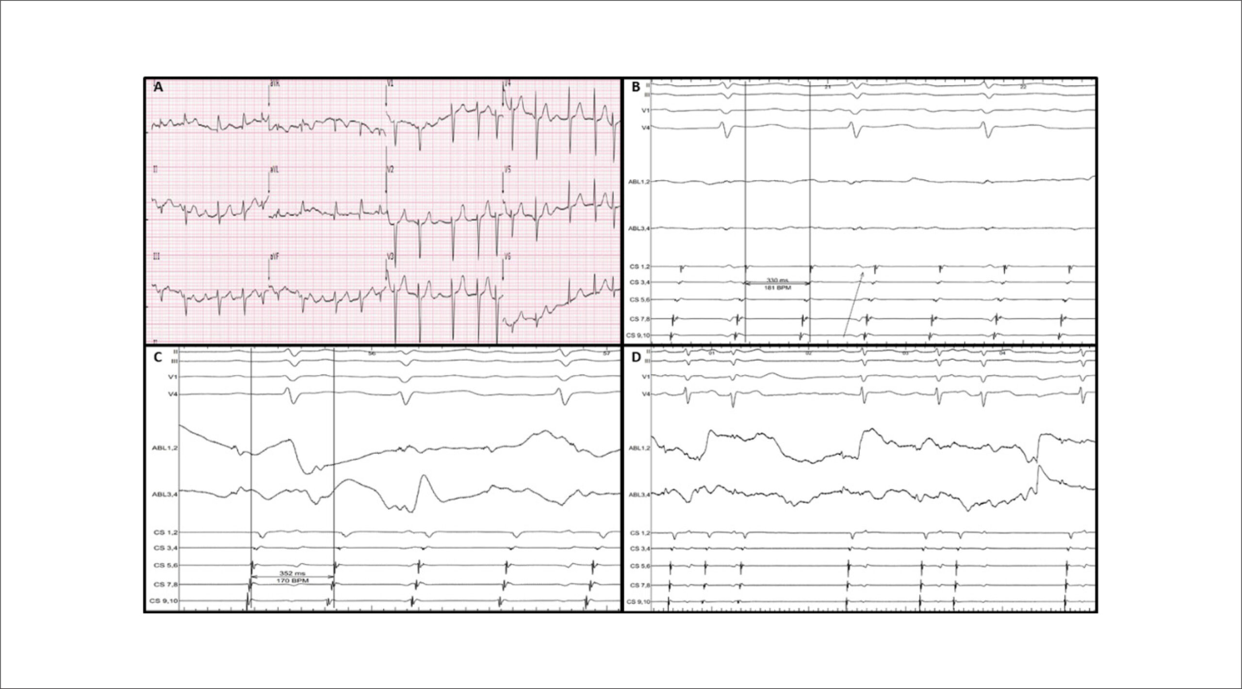
An 86-year-old woman with a previous history of paroxysmal atrial fibrillation was referred for atypical atrial flutter (AFL) ablation. She had never been submitted to catheter ablation before. The transthoracic echocardiogram revealed a left ventricular ejection fraction of 65%, a mildly dilated left atrium (42mm) and moderate mitral regurgitation. The baseline 12-lead ECG exhibited positive F waves in V1 and the lower limb leads, isoelectric F waves in DI and negative F wave in aVL. () Given the earlier concentric activation in the coronary sinus (CS), with a tachycardia cycle length (TCL) of 330ms, () a right atrial (RA) activation map, performed with the PentaRay® catheter, was initially analyzed using the HD Coloring tool (CARTO® 3V7, Biosense Webster, CA, USA). It revealed an earlier activation in the interatrial septum with only 1/3 of the TCL (). A subsequent left atrial (LA) activation map revealed a large area of scar with likely conduction block in almost all of the anterior wall, represented as a white line by the lower threshold Extended Early Meets Late (EEML) feature, and several areas with early local activation time (LAT) points – in the left atrial appendage (LAA), the anterior aspect of the mitral valve (MV) and the roof near the anterior segment of the right superior pulmonary vein (RSPV). It also showed two zones of re-entry as established by the Early Meets Late (EML) feature ( and ). These maps suggested a probable circuit around the right pulmonary veins but did not explain the color activation in the LAA and MV. A new mapping algorithm – Coherent (CARTO® 3V7, Biosense Webster) – unveiled the circuit ( and ). What is the mechanism of this atypical AFL?
[…]