Resident Macrophages Orchestrating Heart Rate
Macrophages and the heartbeat
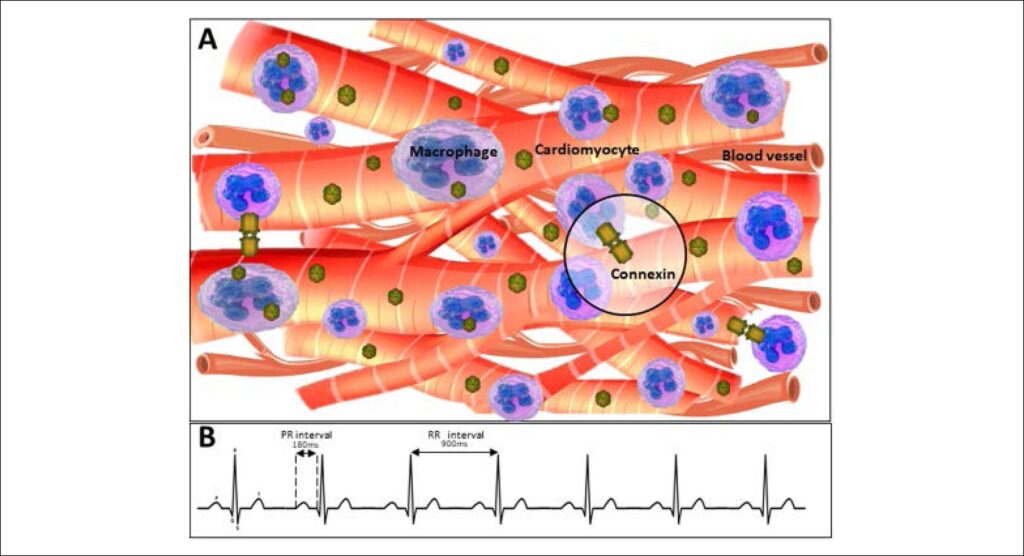
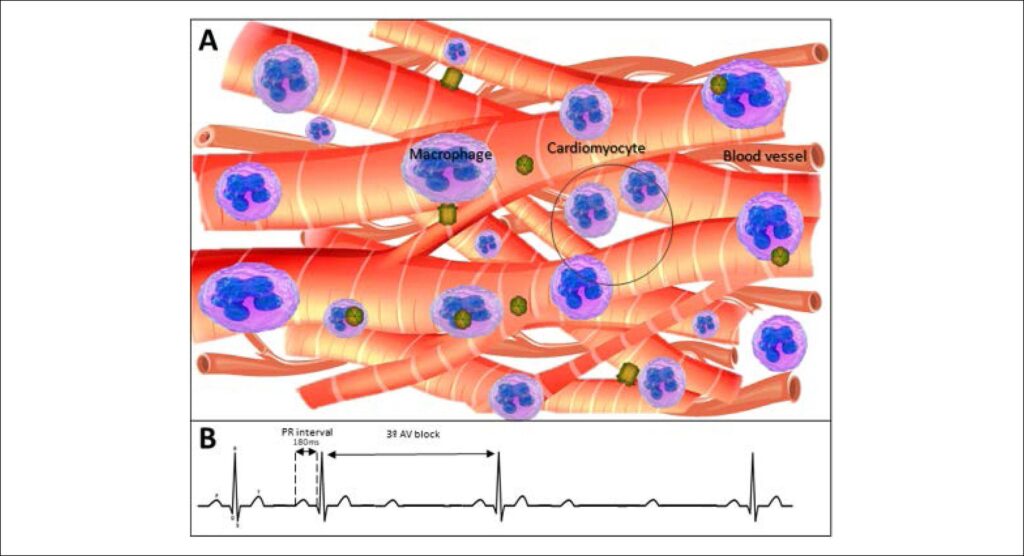
Cardiac function depends on the appropriate moment of contraction in several distinct regions, as well as the heart rate. Hulsmans et al. observed that mice that had their macrophage fauna weakened, had bradycardia and irregular beats. It is known that connexin-43 is predominant in ventricles of humans and that its reduction promotes bradycardia and AV block, thus, in observing specialized cells in non-muscular electrical conduction, they found that macrophages are electrically coupled to cardiomyocytes and that these resident macrophages facilitate electrical conduction through the AV node.
Such conducting cells interleave with macrophages expressing connexin-43 forming additional gap junctions between cardiomyocytes (). The investigators observed that the animals that had a reduction of resident macrophages, besides having bradycardia, had AV blockade of 2nd and 3rd degrees (), whose cause in humans is still unknown. Another intriguing point is that cardiac macrophages have a resting membrane potential of -35 mV on average and depolarize in synchrony with cardiomyocytes. This makes the membrane potential at the rest of the cardiomyocytes more positive and according to the results obtained by computational simulation, accelerate both depolarization and repolarization phases. The cardioprotective role of cardiac resident macrophages can go beyond the modulation of the electrophysiological properties of the coupled cardiomyocytes. The perivascular localization of cardiac macrophages makes them uniquely positioned to interpret systemic signals in the bloodstream.
[…]
Images
Image gallery fullscreen viewer
Comments are moderated by the editorial team before publication. Discussions should be evidence-based and use scientific language. Conflicts of interest should be declared.