Takayasu Arteritis: From Diagnosis to a Life-Threatening Complication
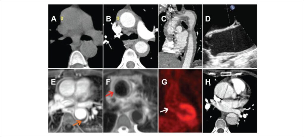
A fifty-two-year-old Caucasian woman was admitted for severe epigastric pain irradiating to the back. Physical examination and electrocardiogram were normal. Laboratory tests showed leucocytosis (11100 cells/uL) and increased levels of C-reactive protein (15.6 mg/dl). Due to the suspicion of acute aortic syndrome (AAS), she underwent computed tomography (CT), which showed a low attenuation circumferential mural thickening of the aorta (43 Hounsfield units (HU)), which enhanced (73 HU) after contrast administration (-), suggestive of aortitis. Transesophageal echocardiogram also revealed thickened thoracic aorta (). Cardiovascular magnetic resonance imaging confirmed the diagnosis of aortitis and excluded intramural hematoma (mural thickening hypointense on T1-weighted images and hyperintense on T2-weighted images), (Figures E-F). Infectious serologies were negative.
[…]
Images
Image gallery fullscreen viewer
Comments are moderated by the editorial team before publication. Discussions should be evidence-based and use scientific language. Conflicts of interest should be declared.