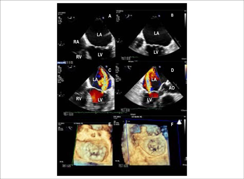
2D-TEE, four-chamber view showing mitral valve prolapse at 0° (A) e 34° (B) rotation of imaging array. C and D. 2D TEE color flow mapping evidencing eccentric mitral regurgitation, directed posteriorly at 0° (C) and 138° (D) rotation of imaging array. E and F. 3D-TEE images showing the mitral valve prolapse of the anterior leaflet (A2) (E, arrow), in a surgical view (from the left atrium, LA) and chordal rupture (F, arrow). TEE: transesophageal echocardiography; RA: right atrium; RV: right ventricule; LV: left ventricule; AO: aorta.
Comments are moderated by the editorial team before publication. Discussions should be evidence-based and use scientific language. Conflicts of interest should be declared.
Comments are moderated by the editorial team before publication. Discussions should be evidence-based and use scientific language. Conflicts of interest should be declared.